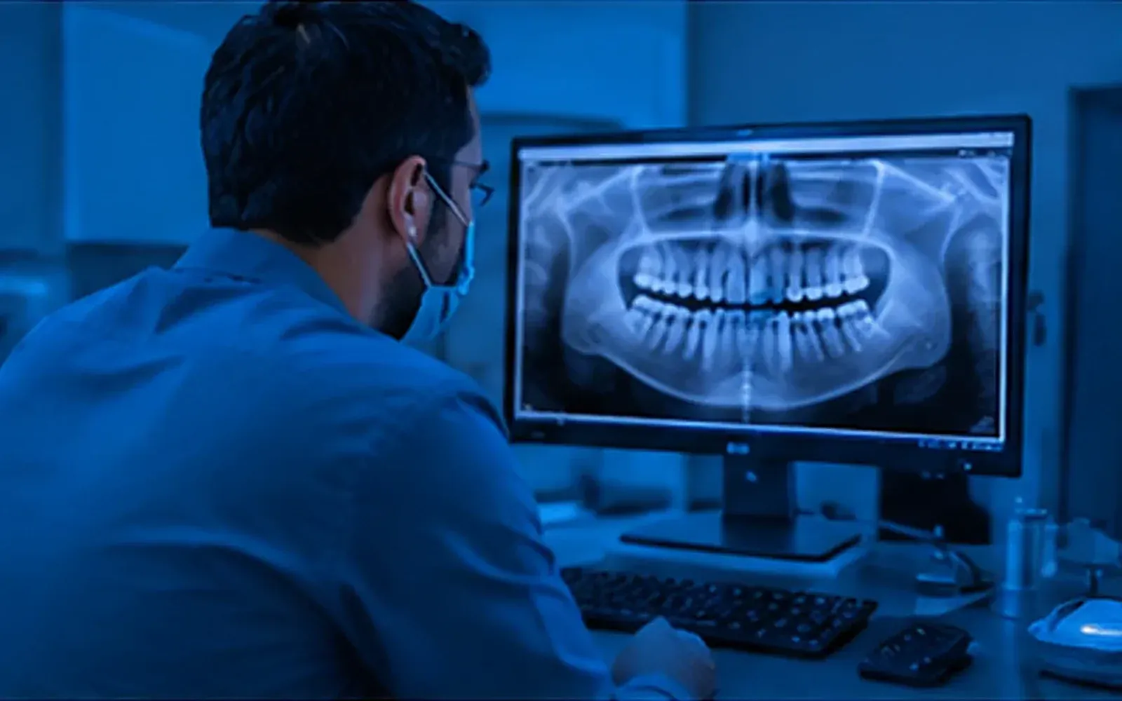
Most clinics have X-rays in three places. Some are in the sensor’s own software on one computer. Some are on a phone, because the intraoral camera was quicker. A few are on a USB stick a patient brought from another clinic. When the dentist needs them during a consultation, someone goes looking.
Imaging software in a clinic system is about putting all of that in one place: the patient’s file. This guide covers what it should do, and what questions to ask.
One place for every kind of image
A dental clinic deals with more image types than most people realise:
- Periapical and bitewing X-rays.
- Panoramic and cephalometric X-rays.
- Intraoral and extraoral photos.
- DICOM files from imaging centres.
- CBCT scans, often as a zip.
- 3D intraoral scans.
Your system should accept all of them in the patient file, sorted by the day they were taken, without anyone converting files first.
BeClarified’s imaging takes X-rays, photos, DICOM, CBCT zips and 3D scans. Drop them onto the patient’s file, or paste a screenshot straight from your sensor software. CBCT zips and 3D scans are recognised automatically. Every file goes through a safety check before it opens, which matters when patients bring files from elsewhere.
Getting photos in from a phone
Phones take excellent intraoral photos, but getting them into the right patient’s file is usually a mess of WhatsApp messages to yourself.
A better way: show a QR code on the computer, scan it with the clinic phone, and every photo taken goes straight into that patient’s images. BeClarified does exactly this, and the code ends when you’re done.
A viewer made for dentists
A generic image viewer isn’t enough for reading X-rays. Look for:
- Zoom, brightness and contrast, plus invert and sharpen, to read a periapical properly.
- Measurement in millimetres. Rulers in pixels mean nothing clinically. A good viewer lets you calibrate: draw over something of known length (a file, an implant) and every ruler on that image then shows millimetres.
- Annotations that sit on top of the image without changing the original file.
- Side-by-side comparison, to show healing or progression over time.
All of these are in the BeClarified viewer. Large CBCT scans open at a lower resolution in the browser, with a download for full detail in dedicated software. 3D scans can be turned, moved and zoomed.
Mounts
A full-mouth series is 14 to 20 images. Reading them as a loose pile is slow. A mount lays them out in their anatomical places, so you read and share them together.
BeClarified includes common mounts (full mouth, bitewings and more), and you can draw your clinic’s own layouts in Settings: set a grid, drag to draw each slot, and name it with its kind and teeth. Built-in layouts can be copied and changed. Archiving a layout doesn’t touch mounts already made with it.
Sharing images with patients
Patients ask for their X-rays: for a second opinion, for a specialist, or just to see them. Burning a CD or printing on paper is slow and expensive.
Share a link instead. In BeClarified you share images by link or QR code for 1, 7 or 30 days, and the patient opens them without signing in. You see when the link was created and how many times it was opened, and you can stop it at any time. Or send the images straight to the patient’s app, where they can zoom and download them. See imaging.
Images in the rest of the workflow
Images are evidence, so they need to travel with the work:
- Attach X-rays to an insurance pre-approval or claim when the company asks for them. See dental insurance claims.
- Attach photos and scans to a lab order, so the lab sees the shade and the preparation.
Once an image is attached to a claim, pre-approval or lab order, BeClarified won’t let it be deleted until it’s taken off there first. That’s a small rule that prevents big problems.
Storage and deletion
Images are big. Ask how much storage is included, what happens when it’s full, and how deletion works. In BeClarified, deleted images go to a trash for 30 days before they’re removed for good, and the file shows how much of your storage you’ve used.
Questions to ask any vendor
- Which file types can I add, including DICOM, CBCT and 3D scans?
- Can I calibrate measurements to millimetres?
- Can I build my own mounts?
- How do I share images with a patient, and for how long?
- Can images be attached to claims and lab orders?
- How much storage is included, and what happens to deleted images?
Frequently asked questions
Does this replace my sensor’s software? No. Keep using your sensor software to capture X-rays, then bring them into the patient file by dropping or pasting them. The patient’s record then has everything in one place.
Can I open CBCT scans in the browser? You can view them, at reduced resolution for large scans. For detailed planning, download the scan and open it in your CBCT software.
What about old images from before? Image files added before Imaging was switched on wait in an “unsorted” list. Give them a kind to bring them in; nothing is moved or lost.
Is sharing images with patients secure? Each link works only for the period you choose, can be stopped at any time, and shows how many times it was opened.